Alexander Nestor-BergmannScientist, mathematician and engineer, working on quantitative research, machine learning and AI.

Alexander Nestor-Bergmann

Scientist, mathematician and engineer, working on quantitative research, machine learning and AI.

A tall research room in oak and plaster. On the left, a framed portrait and a desk with four screens in a fluted timber alcove under the name Unsigned Research. A wall of lit bookshelves fills the far end, with a display of paper covers and a reading table. A whiteboard covered in handwriting hangs on the right, beside a dark panel showing the outlines of cells, and a glowing sheet of cells shaped like an embryo floats above a dark plinth in the middle.

01

About

My background and education.
Alexander Nestor-Bergmann

I’m CTO and co-founder of Unsigned Research, a systematic trading firm. I build and deploy production data pipelines, statistical & machine learning forecasting models and agentic AI systems, using rigorous scientific principles.

I was previously an academic working in mathematics and biophysics. As a Herchel Smith Research Fellow at the University of Cambridge, I studied the theory of active matter and built models of how living tissues take shape. I worked in close collaboration with experimental biologists, using experimental data to inform models and make new predictions.

Education

  1. 2013 – 2017

    PhD in Biological Mathematics

    University of Manchester

  2. 2009 – 2013

    MMath & MPhys, Mathematics and Physics
    University of Manchester

02

Current work

What I do at Unsigned Research.

At Unsigned Research I lead research and engineering. Here are some of the things we do:

  • Time-series forecasting. We build features from noisy, low-signal data, using statistical methods to identify their predictive power. We integrate factors with signal spanning our existing library into our proprietary machine learning models.
  • Risk management. We model and measure risk using factor models and the theory of active portfolio management, creating predictive forecasts to construct our portfolio and explanatory decompositions of realised outcomes.
  • Production infra. We run a scalable cloud-native infrastructure, linked end-to-end: data pipelines, feature engineering, modelling, validation and live execution.
  • AI agents. We design and deploy custom agentic solutions. Our agents work autonomously on research, DevOps and client communication.
  • Deep learning and LLMs. We build, train and fine-tune neural networks and large language models in PyTorch for domain-specific tasks, for example converting text data into numerical features.

Experience

  1. 2022 – present

    Co-founder, CTO & Lead Engineer

    Unsigned Research

    I co-founded the firm and lead the research and engineering of its proprietary systematic investment models.

  2. 2026

    Consultant

    Roche

    I built a full-stack analytics platform for laboratory data, which runs statistical analyses, classifies the results automatically and produces reports.

  3. 2024

    Consultant

    AstraZeneca

    I designed and deployed statistical dashboards that automate the analysis of high-throughput imaging assays.

  4. 2018 – 2022

    Herchel Smith Research Fellow

    University of Cambridge

    I worked across mathematics and biology on the mechanics of cells and how tissues take shape. With experimental collaborators I built deep learning pipelines for microscopy and flow cytometry data, covering cell segmentation, tracking and mechanistic inference.

  5. 2017 – 2018

    Travelling Research Fellow

    University of Queensland

    I modelled and simulated how cells and tissues sense and respond to mechanical force. Our work showed that tissues actively change their material properties in response to mechanical stress.

Full history on LinkedIn

03

Papers

My journal papers, preprints and PhD thesis.

Journal papers

Each preprint is listed with the paper it became.

  1. 2024

    Polarised cell intercalation during Drosophila axis extension is robust to an orthogonal pull by the invaginating mesoderm

    Claire M. Lye, Guy B. Blanchard, Jenny Evans, Alexander Nestor-Bergmann, Bénédicte Sanson

    PLOS Biology 22(4), e3002611

    In the fruit-fly embryo the germband extends along the body axis while the mesoderm, folding in next to it, pulls on it at right angles. We show that the cell rearrangements driving extension carry on despite this pull, which instead slows how fast the cells lengthen along the axis.

    DOI 10.1371/journal.pbio.3002611

  2. 2022

    Adhesion-regulated junction slippage controls cell intercalation dynamics in an Apposed-Cortex Adhesion Model

    Alexander Nestor-Bergmann, Guy B. Blanchard, Nathan Hervieux, Alexander G. Fletcher, Jocelyn Étienne, Bénédicte Sanson

    PLOS Computational Biology 18(1), e1009812

    A model that explicitly represents the apposed cortices of neighbouring cells and the adhesions between them shows that forces at the junctions alone can drive neighbour exchange, and that the turnover of adhesion molecules controls the friction between cells, which in turn sets the material properties of the tissue as a whole.

    DOI 10.1371/journal.pcbi.1009812

  3. 2021

    Generation of anisotropic strain dysregulates wild-type cell division at the interface between host and oncogenic tissue

    Megan Moruzzi, Alexander Nestor-Bergmann, Georgina K. Goddard, Nawseen Tarannum, Keith Brennan, Sarah Woolner

    Current Biology 31(15), 3409–3418.e6

    In frog (Xenopus) embryos, small clusters of cells carrying a cancer-causing form of the Ras gene contract and strain the normal tissue around them, which then divides more often and in altered directions.

    DOI 10.1016/j.cub.2021.05.023

  4. 2019

    Decoupling the Roles of Cell Shape and Mechanical Stress in Orienting and Cueing Epithelial Mitosis

    Alexander Nestor-Bergmann, Georgina A. Stooke-Vaughan, Georgina K. Goddard, Tobias Starborg, Oliver E. Jensen, Sarah Woolner

    Cell Reports 26(8), 2088–2100.e4

    We stretch frog (Xenopus) embryo tissue and use a vertex model to infer the stress in each cell from its shape, which lets us separate the two. The direction a cell divides follows its shape, defined by the corners where three cells meet, while how often cells divide responds to mechanical stress.

    DOI 10.1016/j.celrep.2019.01.102

  5. 2019

    The tricellular vertex-specific adhesion molecule Sidekick facilitates polarised cell intercalation during Drosophila axis extension

    Tara M. Finegan, Nathan Hervieux, Alexander Nestor-Bergmann, Alexander G. Fletcher, Guy B. Blanchard, Bénédicte Sanson

    PLOS Biology 17(12), e3000522

    The adhesion protein Sidekick sits only at the points where three cells meet in fruit-fly epithelia. Without it, cell shapes are abnormal and fewer cells exchange neighbours during axis extension. Our vertex modelling shows how adhesion at these few points can delay neighbour exchange and so change how the whole tissue extends.

    DOI 10.1371/journal.pbio.3000522

  6. 2018

    A Mechanosensitive RhoA Pathway that Protects Epithelia against Acute Tensile Stress

    Bipul R. Acharya, Alexander Nestor-Bergmann, Xuan Liang, Shafali Gupta, Kinga Duszyc, Estelle Gauquelin, Guillermo A. Gomez, Srikanth Budnar, Philippe Marcq, Oliver E. Jensen, Zev Bryant, Alpha S. Yap

    Developmental Cell 47(4), 439–452.e6

    When an epithelium is suddenly put under tension, Myosin VI at cell–cell junctions senses the force and switches on RhoA, which strengthens the junctions, especially where several cells meet, so the sheet holds together.

    DOI 10.1016/j.devcel.2018.09.016

  7. 2018

    Mechanical characterization of disordered and anisotropic cellular monolayers

    Alexander Nestor-Bergmann, Emma Johns, Sarah Woolner, Oliver E. Jensen

    Physical Review E 97(5), 052409

    Starting from a vertex model of individual cells, we derive the emergent mechanical properties of a disordered tissue, such as how it resists stretch and shear. We compare them with stretched frog (Xenopus) tissue, where stretching lines the cells up but stress stays uneven from cell to cell.

    DOI 10.1103/PhysRevE.97.052409

  8. 2018

    Relating cell shape and mechanical stress in a spatially disordered epithelium using a vertex-based model

    Alexander Nestor-Bergmann, Georgina Goddard, Sarah Woolner, Oliver E Jensen

    Mathematical Medicine and Biology: A Journal of the IMA 35, i1–i27

    We use a vertex model to infer the magnitude and direction of the stress in each cell purely from its shape in an image, and apply it to images of frog (Xenopus) embryo tissue.

    DOI 10.1093/imammb/dqx008

  9. 2014

    Force and the spindle: Mechanical cues in mitotic spindle orientation

    Alexander Nestor-Bergmann, Georgina Goddard, Sarah Woolner

    Seminars in Cell & Developmental Biology 34, 133–139

    A review of the evidence that mechanical forces help orient the mitotic spindle, and so the direction of cell division, in single cells and in tissues.

    DOI 10.1016/j.semcdb.2014.07.008

Thesis

  1. 2018

    Relating cell shape, mechanical stress and cell division in epithelial tissues

    Alexander Nestor-Bergmann

    University of Manchester (PhD thesis)

    Combines vertex-model theory of stress in cells and tissues with stretching experiments on frog (Xenopus) tissue to separate the roles of cell shape and mechanical stress in cell division.

Citations and the full record are on Google Scholar.

04

Talks

Talks I've given on my research in tissue mechanics.

  1. Mechanical Characterisation of Disordered and Anisotropic Cellular Monolayers

    British Applied Mathematics Colloquium 2019, University of Bath

  2. Stress and Disorder in a Confluent Epithelium

    Mathematical Biology programme, Institut Mittag-Leffler, Djursholm, Sweden

  3. Shape, Stress and Division in Epithelia

    University of Queensland, Australia

05

Code

Some code I've made public.

  • AI MCP server

    agents-mcp-demo

    A template Model Context Protocol server that gives an AI agent tools for a small PostgreSQL user database.

  • AI research assistant

    ai-deep-research-assistant

    A proof-of-concept research assistant built with Pydantic AI and LangGraph, in which planning, research and synthesis agents answer questions from web searches with citations.

  • Fine-tuned LLM classifier

    hugging-face-llm-classifier

    A framework for fine-tuning Hugging Face transformer models on text classification, with an example that sorts COVID-19 tweets into five sentiment classes using DistilBERT.

  • Transformer from scratch

    pytorch-transformer-demo

    A from-scratch PyTorch implementation of the transformer from "Attention Is All You Need", written up as a tutorial with unit tests.

  • Cell mechanics model

    appcom

    A Python implementation of the Apposed-Cortex Adhesion Model of an epithelium, in which each cell's cortex is an active elastic rod and springs stand for the adhesions between neighbouring cells. Development has moved to a repository at Université Grenoble Alpes.

All repositories on GitHub

06

The maths

Equations from my research on tissue mechanics, next to some standard portfolio theory.

Mechanics of an epithelium

  1. Energy of the cell sheet

    U=∑α=1Nc[12(Aα−1)2+12ΓLα2+12ΛLα]=∑α=1Nc[12(Aα−1)2+12Γ(Lα−L0)2]−NcΛ28Γ,L0=−Λ2Γ\begin{aligned}U &= \sum_{\alpha=1}^{N_c} \Big[ \tfrac{1}{2}\left(A_\alpha - 1\right)^2 + \tfrac{1}{2}\Gamma L_\alpha^2 + \tfrac{1}{2}\Lambda L_\alpha \Big] \\[0.35em] &= \sum_{\alpha=1}^{N_c} \Big[ \tfrac{1}{2}\left(A_\alpha - 1\right)^2 + \tfrac{1}{2}\Gamma\left(L_\alpha - L_0\right)^2 \Big] - \frac{N_c\Lambda^2}{8\Gamma}, \\[0.35em] L_0 &= -\frac{\Lambda}{2\Gamma}\end{aligned}

    Each of the NcN_c polygonal cells α\alpha, with area AαA_\alpha and perimeter LαL_\alpha, pays an energy for changing its area from the preferred value and for stretching its cortex away from a target perimeter L0L_0 (positive when Λ<0\Lambda < 0); lengths are measured in units of the square root of the preferred area.Nestor-Bergmann et al., Math. Med. Biol. 35, i1–i27 (2018), §3.2.

  2. The two parameters

    Γ=Γ~K~A~0,Λ=Λ~K~A~03/2\begin{aligned}\Gamma &= \frac{\tilde{\Gamma}}{\tilde{K}\tilde{A}_0}, \qquad \\[0.35em] \Lambda &= \frac{\tilde{\Lambda}}{\tilde{K}\tilde{A}_0^{3/2}}\end{aligned}

    Only two numbers set the behaviour, the cortical contractility Γ\Gamma and the line tension Λ\Lambda, each measured against the cell’s resistance to area change K~\tilde{K} and its preferred area A~0\tilde{A}_0 (tildes mark dimensional quantities).Nestor-Bergmann et al., Math. Med. Biol. 35, i1–i27 (2018), §3.2.

  3. Pressure and tension of a cell

    Pα≡Aα−1,Tα≡Γ(Lα−L0)P_\alpha \equiv A_\alpha - 1, \qquad T_\alpha \equiv \Gamma\left(L_\alpha - L_0\right)

    In the paper’s sign convention, PαP_\alpha is positive when a cell is larger than its preferred area, and TαT_\alpha is positive when its perimeter is longer than L0L_0. In both cases the cell pulls inward on its vertices, so positive PαP_\alpha means tension, not compression.Nestor-Bergmann et al., Math. Med. Biol. 35, i1–i27 (2018), §3.2.

  4. Force at a vertex

    fαi=−Pα pαi+Tα qαi,pαi=12(Rαi+1−Rαi−1)×z^,qαi=t^αi−t^αi−1\begin{aligned}\mathbf{f}_\alpha^i &= -P_\alpha\,\mathbf{p}_\alpha^i + T_\alpha\,\mathbf{q}_\alpha^i, \\[0.35em] \mathbf{p}_\alpha^i &= \tfrac{1}{2}\left(\mathbf{R}_\alpha^{i+1} - \mathbf{R}_\alpha^{i-1}\right)\times\hat{\mathbf{z}}, \\[0.35em] \mathbf{q}_\alpha^i &= \hat{\mathbf{t}}_\alpha^{i} - \hat{\mathbf{t}}_\alpha^{i-1}\end{aligned}

    The force cell α\alpha exerts on its vertex ii is minus the gradient of its energy, with a pressure part along the outward direction pαi\mathbf{p}_\alpha^i and a tension part along the inward direction qαi\mathbf{q}_\alpha^i; vertices are numbered anticlockwise, Rαi\mathbf{R}_\alpha^i runs from the cell’s centre (the mean of its vertices) to vertex ii, and t^αi\hat{\mathbf{t}}_\alpha^i is the unit vector along the edge from vertex ii to i+1i+1.Nestor-Bergmann et al., Math. Med. Biol. 35, i1–i27 (2018), §3.2.

  5. Stress in a cell

    σα=−Pαeff I+TαJα−12S˙α,Pαeff=Pα+TαLα2Aα,Jα=1Aα(12LαI−∑i=0Zα−1lαi t^αi⊗t^αi)\begin{aligned}\boldsymbol{\sigma}_\alpha &= -P_\alpha^{\text{eff}}\,\mathsf{I} + T_\alpha \mathsf{J}_\alpha - \tfrac{1}{2}\dot{\mathsf{S}}_\alpha, \\[0.35em] P_\alpha^{\text{eff}} &= P_\alpha + \frac{T_\alpha L_\alpha}{2A_\alpha}, \\[0.35em] \mathsf{J}_\alpha &= \frac{1}{A_\alpha}\Big( \tfrac{1}{2}L_\alpha\mathsf{I} - \sum_{i=0}^{Z_\alpha-1} l_\alpha^i\,\hat{\mathbf{t}}_\alpha^i \otimes \hat{\mathbf{t}}_\alpha^i \Big)\end{aligned}

    The first moment of the vertex forces over a cell, divided by its area, gives its stress: an isotropic effective pressure that adds the cortex tension in Young–Laplace form to the bulk pressure, a traceless part Jα\mathsf{J}_\alpha set by the cell’s ZαZ_\alpha edges (edge ii has length lαil_\alpha^i), and, out of equilibrium, a viscous part from the rate of change of the shape tensor Sα=(1/Zα)∑iRαi⊗Rαi\mathsf{S}_\alpha = (1/Z_\alpha)\sum_i \mathbf{R}_\alpha^i \otimes \mathbf{R}_\alpha^i. Signs follow the paper: a cell under net tension has Pαeff>0P_\alpha^{\text{eff}} > 0.Nestor-Bergmann et al., Math. Med. Biol. 35, i1–i27 (2018), §3.3.

  6. Balance across the sheet

    ∑α=1NcAαPαeff=A Pext,∑α=1NcAαTαJα=0,A=∑α=1NcAα\begin{aligned}\sum_{\alpha=1}^{N_c} A_\alpha P_\alpha^{\text{eff}} &= A\,P_{\text{ext}}, \\[0.35em] \sum_{\alpha=1}^{N_c} A_\alpha T_\alpha \mathsf{J}_\alpha &= \mathsf{0}, \\[0.35em] A &= \sum_{\alpha=1}^{N_c} A_\alpha\end{aligned}

    At equilibrium some cells are under tension and some under compression, but their effective pressures, averaged by area, must equal the pressure PextP_{\text{ext}} applied around the edge of the sheet (Pext>0P_{\text{ext}} > 0 pulls it outward), and their area-weighted shear stresses must cancel.Nestor-Bergmann et al., Math. Med. Biol. 35, i1–i27 (2018), §3.5.

  7. Bulk modulus of a disordered sheet

    K=∑α=1NcAα2A[2Aα+ΓL0Lα2Aα]→hexagons  K=Aα−Λ μ68Aα,μ6=223\begin{aligned}K &= \sum_{\alpha=1}^{N_c} \frac{A_\alpha}{2A}\left[ 2A_\alpha + \frac{\Gamma L_0 L_\alpha}{2A_\alpha} \right] \\[0.35em] &\xrightarrow{\text{hexagons}}\; \\[0.35em] K &= A_\alpha - \frac{\Lambda\,\mu_6}{8\sqrt{A_\alpha}}, \\[0.35em] \mu_6 &= 2\sqrt{2\sqrt{3}}\end{aligned}

    If every cell of an unloaded sheet is stretched uniformly by the same small amount, its resistance to area change is a sum over the cells; for identical hexagons, where uniform stretching is exact, it reduces to a closed form (μ6\mu_6 is the perimeter of a regular hexagon divided by the square root of its area).Nestor-Bergmann et al., Math. Med. Biol. 35, i1–i27 (2018), §3.6.

Portfolio theory

  1. Linear factor model

    r=Xb+u,r,u∈RN,  X∈RN×K,  b∈RK\begin{aligned}\mathbf{r} &= \mathbf{X}\mathbf{b} + \mathbf{u}, \\[0.35em] &\mathbf{r},\mathbf{u} \in \mathbb{R}^{N},\; \\[0.35em] &\mathbf{X} \in \mathbb{R}^{N\times K},\; \\[0.35em] &\mathbf{b} \in \mathbb{R}^{K}\end{aligned}

    The excess returns r\mathbf{r} of NN assets are explained by their exposures X\mathbf{X} to KK common factors, times the factor returns b\mathbf{b}, plus a specific return u\mathbf{u} for each asset that is uncorrelated with the factors and with the other assets’ specific returns.

  2. Factor risk model

    V=XFX⊤+Δ,σP2=h⊤Vh=x⊤Fx+h⊤Δh,x=X⊤h\begin{aligned}\mathbf{V} &= \mathbf{X}\mathbf{F}\mathbf{X}^{\top} + \boldsymbol{\Delta}, \\[0.35em] \sigma_P^2 &= \mathbf{h}^{\top}\mathbf{V}\mathbf{h} = \mathbf{x}^{\top}\mathbf{F}\mathbf{x} + \mathbf{h}^{\top}\boldsymbol{\Delta}\mathbf{h}, \\[0.35em] \mathbf{x} &= \mathbf{X}^{\top}\mathbf{h}\end{aligned}

    The N×NN \times N covariance of returns is built from the K×KK \times K factor covariance F\mathbf{F} and the diagonal matrix of specific variances Δ\boldsymbol{\Delta}, so a portfolio with holdings h\mathbf{h} has a factor risk set by its exposures x\mathbf{x} and a specific risk.

  3. Fundamental law of active management

    IR≈IC BR\mathrm{IR} \approx \mathrm{IC}\,\sqrt{\mathrm{BR}}

    The information ratio, residual return per unit of residual risk, is roughly the skill of each forecast, measured as its correlation with outcomes, times the square root of the number of independent forecasts made each year; it assumes independent forecasts of equal skill and no constraints on the portfolio.

  4. Mean-variance optimal active portfolio

    hA∗=arg⁡max⁡hA{α⊤hA−λA hA⊤VhA}=12λA V−1α,IR∗=α⊤V−1α,ω∗=IR∗2λA\begin{aligned}\mathbf{h}_A^{*} &= \arg\max_{\mathbf{h}_A}\Big\{ \boldsymbol{\alpha}^{\top}\mathbf{h}_A - \lambda_A\,\mathbf{h}_A^{\top}\mathbf{V}\mathbf{h}_A \Big\} \\[0.35em] &= \frac{1}{2\lambda_A}\,\mathbf{V}^{-1}\boldsymbol{\alpha}, \\[0.35em] \mathrm{IR}^{*} &= \sqrt{\boldsymbol{\alpha}^{\top}\mathbf{V}^{-1}\boldsymbol{\alpha}}, \\[0.35em] \omega^{*} &= \frac{\mathrm{IR}^{*}}{2\lambda_A}\end{aligned}

    With alpha forecasts α\boldsymbol{\alpha} and risk aversion λA\lambda_A, and no constraints, the best active holdings are proportional to V−1α\mathbf{V}^{-1}\boldsymbol{\alpha}; the ex-ante information ratio of that portfolio depends only on the forecasts and the risk model, and λA\lambda_A only sets how much active risk ω\omega is taken.

Standard results from published portfolio theory.

07

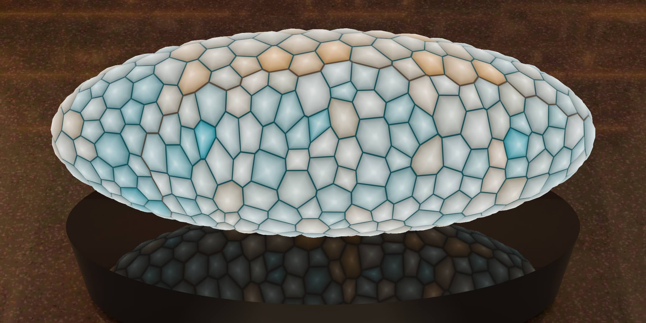
Tissue simulation

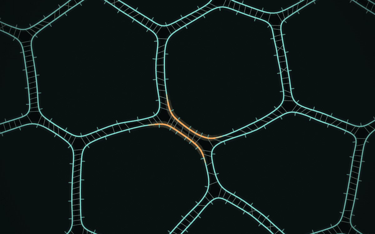
Cell junctions in the outlined region are more contractile, forcing the cells to swap neighbours, so the whole tissue narrows along one axis and extends along the other.

This is a simulation of a fundamental process in development, in which a tissue restructures itself and changes shape using only forces generated within it. We model the cells of the embryo with a vertex model, which represents each cell as a polygon with a preferred area and a contractile perimeter. In the outlined region, the vertical junctions are more contractile, because of a genetic patterning of myosin II, the motor protein that makes cells contract. These junctions shrink until four cells meet at a point, and a new junction then grows at right angles to the old one. As cells exchange neighbours like this, the sheet narrows in one direction and lengthens in the other, as the germband (the strip of tissue that becomes the fly’s body segments) does when the embryo’s body axis extends. Drag the embryo to turn it, and click or tap a cell to remove it.

08

Cell simulation

A simulation of cell intercalation via junction contraction. Each cell has its own cortex, held to its neighbours by adhesions.

This is a small version of the Apposed-Cortex Adhesion Model, from my 2022 paper on how cells in a sheet exchange neighbours. Most models of epithelia draw the boundary between two cells as a single shared edge. In this one every cell has its own cortex, and the cortices of neighbouring cells are held together by adhesions that bind and unbind.

In the model, when the cortices along a junction contract, the junction shortens until four cells meet. The adhesions let the two cortices slip past each other, a new junction opens at right angles, and the cells have swapped neighbours. Click or tap a junction to make it the one that contracts.

What runs here is simplified so that it works in a browser and can repeat. The paper has the full model, and the code is public.

09

Interests

The subjects I work in.

  • Statistics

    Statistical modelling and inference for noisy data.

  • Machine Learning

    Forecasting models, from linear models to deep networks.

  • Agentic AI and LLMs

    Building AI agents, and training and fine-tuning language models.

  • Quantitative Research

    Finding and testing predictive signals in financial markets.

  • Production Trading Systems

    The infrastructure that runs our models and trades live.

  • Computational Biology

    Mathematical models of how cells and tissues take shape.